
Quantitative Morphological Analysis of Various Block Copolymer Micelles
Ryan Pyo
Co-Presenters: Juan Sinta
College: The Dorothy and George Hennings College of Science, Mathematics and Technology
Major: Biology
Faculty Research Mentor: Brian Ree
Abstract:
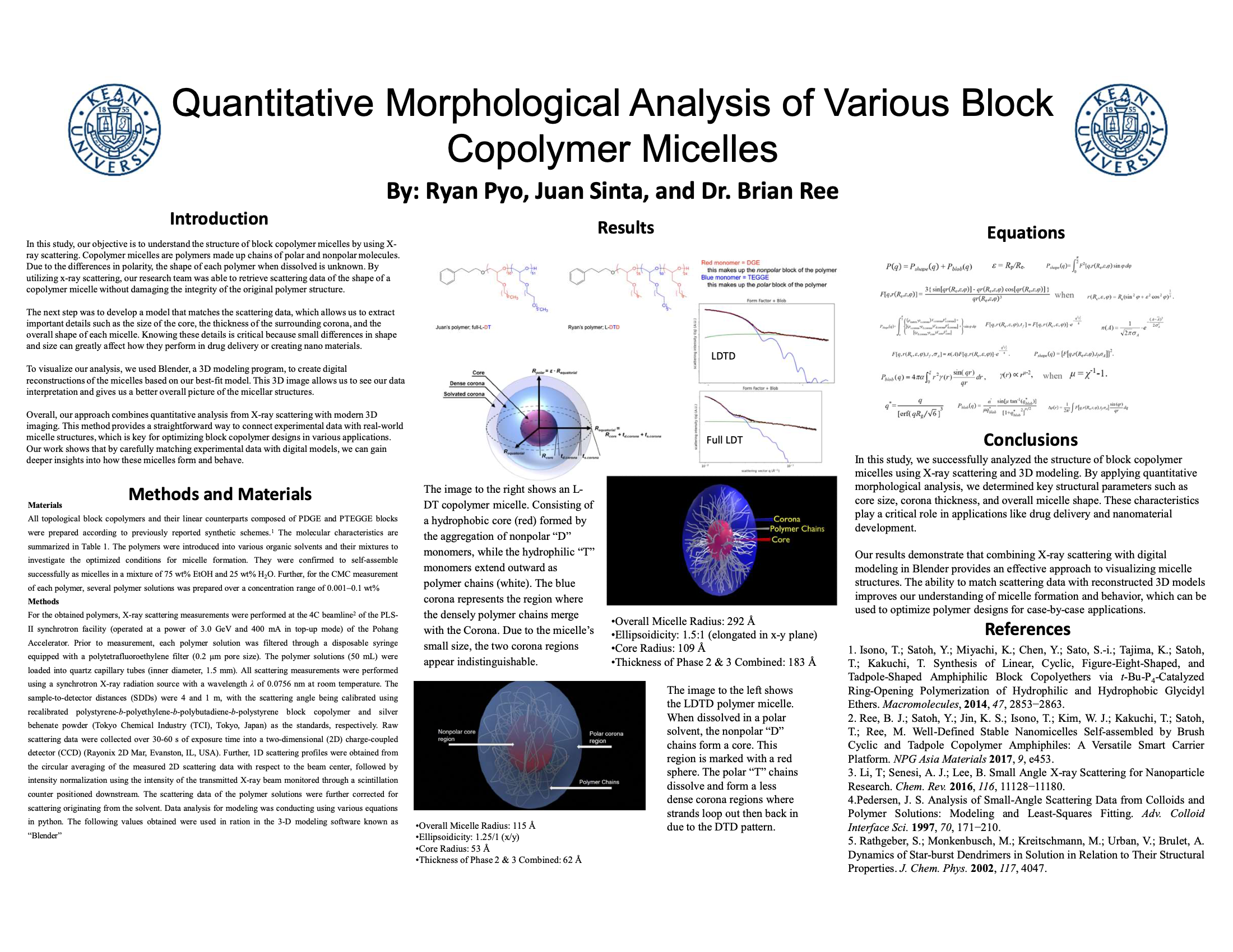
In this study, our objective is to understand the structure of block copolymer micelles by using X-ray scattering. Copolymer micelles are polymers made up chains of polar and nonpolar molecules. Due to the differences in polarity, the shape of each polymer when dissolved is unknown. By utilizing x-ray scattering, our research team was able to retrieve scattering data of the shape of a copolymer micelle without damaging the integrity of the original polymer structure.The next step was to develop a model that matches the scattering data, which allows us to extract important details such as the size of the core, the thickness of the surrounding corona, and the overall shape of each micelle. Knowing these details is critical because small differences in shape and size can greatly affect how they perform in drug delivery or creating nano materials.To visualize our analysis, we used Blender, a 3D modeling program, to create digital reconstructions of the micelles based on our best-fit model. This 3D image allows us to see our data interpretation and gives us a better overall picture of the micellar structures.Overall, our approach combines quantitative analysis from X-ray scattering with modern 3D imaging. This method provides a straightforward way to connect experimental data with real-world micelle structures, which is key for optimizing block copolymer designs in various applications. Our work shows that by carefully matching experimental data with digital models, we can gain deeper insights into how these micelles form and behave.